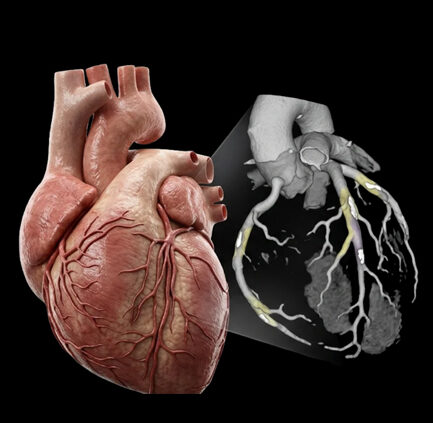


Medical imaging is moving beyond the race for sharper pictures.
The next generation of MRI, CT and ultrasound is being shaped by a different set of priorities: faster examinations, lower radiation exposure, richer diagnostic information, smaller equipment and artificial intelligence (AI) that can assist throughout the imaging workflow.
Some of these changes are already reaching clinical practice. Others remain under evaluation. The most important developments are happening in three areas:
| Modality | Main innovation | What it could change |
| MRI | AI acceleration and portable low-field systems | Faster scans and imaging outside traditional MRI suites |
| CT | Photon-counting detectors | Higher resolution, spectral information and potentially better dose efficiency |
| Ultrasound | AI + handheld systems | More accessible point-of-care imaging |
The direction is clear: medical imaging is becoming faster, more quantitative and more portable, while AI is moving deeper into the imaging process.
MRI: Can AI make a traditionally slow scan faster?
MRI provides excellent soft-tissue imaging without ionizing radiation, but conventional examinations can be lengthy. Patient movement can also reduce image quality.
AI is now being used to address both problems.
A review published in Radiology examined the clinical use of AI-accelerated MRI and found that the technology is already entering routine practice. AI can reconstruct images from less or faster-acquired data, potentially shortening examinations and reducing motion-related problems.
What AI-accelerated MRI is trying to achieve
| Conventional challenge | AI-assisted approach | Potential benefit |
| Long scan times | Faster acquisition + AI reconstruction | Shorter examinations |
| Patient movement | Shorter sequences | Fewer motion artifacts |
| Scanner capacity | Higher throughput | More examinations per scanner |
| Patient discomfort | Reduced time inside scanner | Better experience |
But speed comes with a critical question: Does the reconstructed image preserve the information a radiologist needs?
The RSNA review highlights concerns around AI-generated image changes, including the possibility that lesions could become less conspicuous or, in some circumstances, be artificially introduced. The authors emphasize that the appropriate level of acceleration still needs careful clinical validation.
That makes AI-accelerated MRI less about simply “making MRI faster” and more about finding the point where speed and diagnostic reliability remain balanced.
MRI is also getting smaller
The other major MRI trend is moving in the opposite direction from the industry’s historical focus on increasingly powerful scanners.
Researchers are developing low-field and ultra-low-field MRI systems that can be smaller and potentially portable.
A recent prospective outpatient study involving 125 patients compared portable ultra-low-field MRI with standard MRI for common neurological indications.
The results were notable:
| Finding | Result |
| Participants | 125 |
| Concordance on blinded review | 92% |
| Concordance after clinically informed review | 98% |
| Patients preferring portable MRI | 61% |
| Patients preferring standard MRI | 14% |
The researchers concluded that portable MRI showed high clinical concordance with standard MRI for identifying the presence or absence of structural brain abnormalities and was strongly preferred by patients.
However, portable MRI did not match conventional MRI in every situation. Subtle findings can remain difficult to detect because ultra-low-field systems have lower spatial resolution and fewer specialized sequences.
That suggests a more realistic future:
Portable MRI is more likely to expand access to MRI than to replace high-field MRI altogether.
It could be particularly useful in outpatient neurology, intensive care, emergency settings and locations where installing a conventional MRI scanner is difficult.
CT: Photon counting could change what CT can see
CT is undergoing a more fundamental hardware change.
Photon-counting CT (PCCT) replaces conventional energy-integrating detectors with photon-counting detectors that measure individual X-ray photons and their energy.
That allows the system to capture additional information from the same scan.
The potential advantages include:
- higher spatial resolution
- improved contrast resolution
- spectral information
- lower electronic noise
- improved dose efficiency
- material-specific imaging
The technology is no longer confined to early research.
A 2026 Radiology review from RSNA examined 458 high-impact peer-reviewed clinical publications on photon-counting CT. The studies covered cardiac, thoracic, neurovascular, abdominal, musculoskeletal and pediatric imaging.
Photon-counting CT: where the evidence is emerging
| Area | Reported opportunity |
| Cardiac imaging | Better visualization of coronary arteries, calcification and stents |
| Thoracic imaging | Improved visualization of small structures |
| Neurovascular imaging | High-resolution and spectral information |
| Abdominal imaging | Lesion detection and material characterization |
| Musculoskeletal imaging | Better visualization of subtle bone structures |
| Pediatric imaging | Potential dose-efficiency advantages |
The first commercially available whole-body PCCT system received FDA clearance in 2021. The technology has since expanded into multiple clinical applications.
More information from the same CT scan
One of the most important advantages of photon counting is not simply sharper images.
It is additional information.
Conventional CT primarily provides anatomical information. Photon-counting CT can also generate spectral reconstructions and material maps.
That means CT can increasingly move from:
“Where is the abnormality?”
toward:
“What is the abnormality made of?”
The potential applications are significant.
For example, a recent Radiology study found that photon-counting CT could quantify liver fat, with excellent agreement with MRI-based proton-density fat fraction measurements. In 125 patients who underwent both examinations, the reported intraclass correlation coefficient was 0.91.
That is an interesting direction because information that traditionally required another examination could potentially be extracted from a CT scan that has already been performed.
Resolution is also changing
The first whole-body photon-counting CT system, Siemens Healthineers’ NAEOTOM Alpha, has reported spatial resolution of approximately 0.11 mm at isocenter and temporal resolution of up to 66 milliseconds under specified conditions.
Clinical research is beginning to show what that can mean.
In an early human coronary CT study, 14 participants underwent both photon-counting CT and conventional dual-layer CT angiography. Radiologists rated overall image quality and diagnostic confidence higher with PCCT. For example, image-quality improvement was reported in 100% of calcification assessments and 92% of stent assessments.
More recent research has also found advantages for detecting small lesions. A 2025 Radiology study reported lesion-detection sensitivity of 82% with PCCT versus 78% with conventional energy-integrating detector CT. For subcentimeter lesions, sensitivity was 74% versus 67%.
These are not universal improvements across every CT examination, but they show why photon counting is attracting attention.
What about radiation dose?
Dose efficiency is another major reason for interest in PCCT.
The 2026 RSNA review reports substantial dose reductions in selected clinical applications, including studies reporting more than 70% reductions in some pediatric temporal-bone and musculoskeletal examinations while maintaining or improving image quality.
But this needs context.
A 70% reduction should not be presented as the expected dose reduction for every photon-counting CT scan.
Performance depends on the scanner, protocol, patient and clinical task.
The stronger conclusion is that photon-counting technology creates new opportunities to improve image quality and dose efficiency at the same time.
The RSNA review also identifies protocol standardization and further diagnostic-performance studies as important remaining gaps.
Ultrasound: The handheld revolution meets AI
Ultrasound has an advantage that MRI and CT cannot easily match.
It can be performed at the bedside using relatively small equipment, produces images in real time and does not use ionizing radiation.
Its major weakness is operator dependence.
AI could help address that limitation.
Researchers are developing systems that can assist with image acquisition, image quality, measurements and interpretation.
A multicenter study evaluating AI-assisted handheld ultrasound for cardiac assessment included 200 patients. The AI system achieved 85% sensitivity and 81% specificity for detecting left ventricular ejection fraction below 50%.
The potential evolution of ultrasound
| Today | Emerging direction |
| Clinician positions probe | AI may assist with positioning |
| Clinician selects images | AI can identify useful views |
| Manual measurements | Automated measurements |
| Expert interpretation | AI-assisted interpretation |
| Large ultrasound systems | Increasing use of handheld devices |
This could make ultrasound more useful outside specialist imaging departments.
The long-term opportunity is therefore not simply AI reading ultrasound images.
It is AI helping clinicians acquire better ultrasound images in the first place.
The real change: AI is moving upstream
This may be the most important trend across all three technologies.
AI is no longer limited to finding abnormalities after an examination.
It is increasingly being considered across the entire imaging workflow:
Acquisition → reconstruction → quality control → segmentation → measurement → detection → interpretation
The difference is significant.
Earlier model
Patient → Scanner → Image → Radiologist
Emerging model
Patient → Optimized acquisition → AI reconstruction → Quality assessment → Quantification → AI-assisted interpretation → Radiologist
That means the competitive landscape is changing as well.
Imaging companies are no longer competing only on scanner hardware. They are increasingly competing on detectors, reconstruction algorithms, AI models, workflow software, data and clinical validation.
But innovation still has to prove itself
The rapid development of imaging AI and new hardware does not mean every new system is ready for widespread use.
Three questions remain critical.
- Does it improve clinical decisions?
Better-looking images are not enough. The technology must demonstrate meaningful diagnostic or workflow benefits.
- Is it reliable across patients?
AI performance can change across populations, scanners and clinical settings.
- Can it be implemented consistently?
The 2026 RSNA review of PCCT specifically identified protocol standardization as an important remaining challenge for larger multicenter studies.
These issues will become increasingly important as imaging AI becomes more deeply integrated into clinical workflows.
Where is medical imaging heading?
The three technologies are taking different paths.
| Technology | Direction of travel |
| MRI | Faster, AI-accelerated and increasingly portable |
| CT | Higher resolution, photon counting and spectral imaging |
| Ultrasound | Handheld, AI-assisted and increasingly point-of-care |
| AI | Moving from interpretation into acquisition and reconstruction |
| Healthcare delivery | More remote, decentralized and data-driven |
The biggest change may not be a single new scanner.
It may be the combination of better hardware, AI, quantitative imaging and portability.
MRI could become faster and available in more locations. CT could provide more information from a single examination while improving dose efficiency. Ultrasound could become easier for non-specialists to acquire and interpret.
The future of medical imaging is therefore not simply about seeing more.
It is about getting clinically useful information faster, extracting more information from every scan and making that information available to more patients.
For imaging companies and IP teams, that shift also creates a new competitive battlefield: the valuable innovation may increasingly sit at the intersection of hardware, AI, reconstruction, data and clinical workflow.
Sources
- https://pubs.rsna.org/doi/10.1148/radiol.251496
- https://pubs.rsna.org/doi/10.1148/radiol.243819
- https://pubs.rsna.org/doi/10.1148/radiol.253674
- https://pmc.ncbi.nlm.nih.gov/articles/PMC13495659/
- https://pubs.rsna.org/doi/full/10.1148/radiol.240662
- https://pubs.rsna.org/doi/10.1148/radiol.211780
- https://pubs.rsna.org/doi/10.1148/radiol.243329
- https://pubs.rsna.org/doi/10.1148/radiol.250480
- https://pubs.rsna.org/doi/10.1148/ryct.250072
- https://www.fda.gov/medical-devices/digital-health-center-excellence/artificial-intelligence-enabled-medical-devices
- https://www.fda.gov/medical-devices/artificial-intelligence-enabled-medical-devices/list-artificial-intelligence-enabled-medical-devices
- https://www.who.int/activities/strengthening-medical-imaging/strengthening-medical-imaging